FIG 4.
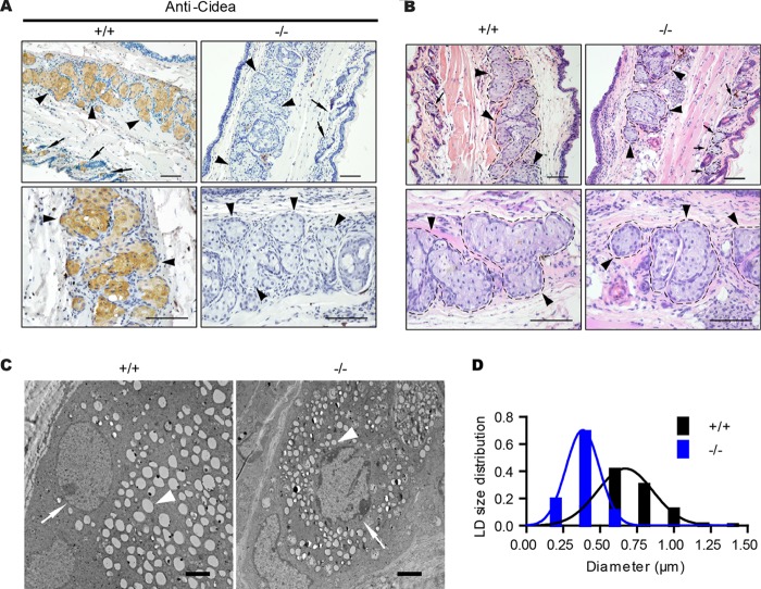
Meibomian-gland-specific expression of Cidea and reduced LD size in the meibomian gland of Cidea−/− mice. (A) Immunohistochemical (IHC) staining of paraffin sections of eyelid from wild-type (+/+) and Cidea−/− (−/−) mice with Cidea antibody. Scale bar, 100 μm. Meibomian glands are indicated by arrowheads, and sebaceous glands are indicated by arrows. (B) H&E staining of eyelid sections from wild-type (+/+) and Cidea−/− (−/−) mice. Meibomian glands are indicated by arrowheads, and sebaceous glands are indicated by arrows. Scale bar, 100 μm. (C) Electron microscopic (EM) images of eyelid sections from wild-type (+/+) and Cidea−/− (−/−) mice. Nuclei are indicated by arrows, and LDs are indicated by arrowheads. Scale bar, 2 μm. (D) Lipid droplet (LD) size distribution. n = 3 for each genotype. Diameters of over 800 LDs for each genotype were measured.

