
FIG 7.
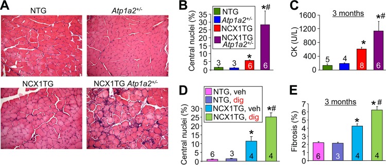
Haploinsufficiency of Atp1a2 or digoxin exacerbates MD in NCX1 TG mice. (A) Representative H&E-stained histological images of the quadriceps (×200 magnification) of NTG, Atp1a2+/−, NCX1 TG, and NCX1 TG Atp1a2+/− mice at 3 months of age. (B) Percentages of fibers with centrally localized nuclei at 3 months of age from the same muscles shown in panel A. *, P < 0.05 versus WT mice; #, P < 0.05 versus NCX1 TG mice. (C) Serum CK levels measured in the indicated groups at 3 months of age. *, P < 0.05 versus WT mice; #, P < 0.05 versus NCX1 TG mice. (D) Percent centrally localized nuclei in histological sections from quadriceps muscles of the groups shown. *, P < 0.05 versus NTG-vehicle-treated (veh) mice; #, P < 0.05 versus NCX1 TG-digoxin-treated (dig) mice. (E) Percent fibrosis measured by Masson's trichrome in histological sections of quadriceps muscles of the groups shown. *, P < 0.05 versus NTG-vehicle treated mice; #, P < 0.05 versus NCX1 TG-digoxin-treated mice. The error bars indicate SEM. Numbers in the bars represent the number of mice analyzed.