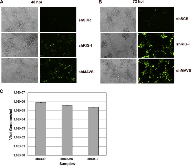
FIG 2.
KSHV infection of control and knockdown cell lines was monitored for GFP expression by fluorescence microscopy for 48 or 72 h. Control cells and MAVS-depleted and RIG-I-depleted stable knockdown 293 cells were infected for 48 h (A) or 72 h (B). GFP expression was significantly increased in both MAVS-depleted and RIG-I-depleted cells compared to the scrambled control. (C) Quantitation of viral genomes at 2 h postinfection (30 min postspinoculation). There was no significant difference in viral genomes detected between the shMAVS and shRIG-I cells compared to shSCR cells at this time point.

