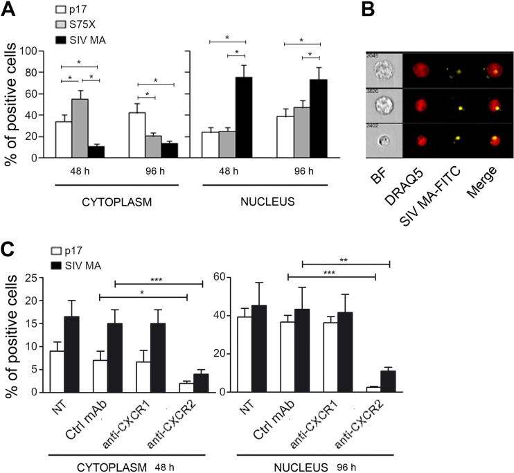
FIG 4.
SIV MA efficiently internalizes into HBL-1 cells and human primary B cells and rapidly translocates into the nucleus. (A) HBL-1 cells were treated for 48 and 96 h with p17-APC, S75X-APC, and SIV MA-FITC at a final concentration of 0.5 μg/106 cells. A total of 2 × 10⁴ HBL-1 cells were acquired with ImageStream X and analyzed using IDEAS software. Nuclear localization of p17, S75X, and SIV MA was calculated as similarity score between labeled protein and nuclear dye intensities. The graph represents a mean of four independent experiments. After both 48 and 96 h of treatment, the SIV MA nuclear localization was significantly higher than the p17 and S75X nuclear localization (*, P < 0.05). The statistical significance was calculated by using Student two-tailed t test. (B) HBL-1 were cultured for 48 h in the presence of SIV MA-FITC and then labeled with the vital nuclear dye DRAQ5. SIV MA-FITC localized almost completely into the nucleus, as demonstrated by multispectral imaging analysis showing SIV MA-FITC/DRAQ5 colocalization. BF, bright field. The data are representative from one representative experiment. (C) Human primary B cells were pretreated with 5 μg of MAb to CXCR1, MAb to CXCR2, or Ctrl MAb/ml for 1 h at room temperature and then treated for 48 and 96 h with p17-APC and SIV MA-FITC (0.5 μg/106 cells). Cells were acquired with ImageStream X and analyzed with IDEAS software. Nuclear localization of p17 and SIV MA was calculated as the similarity score between labeled protein and nuclear dye intensities. The graph represents the mean of two independent experiments. Internalization and nuclear localization of p17 and SIV MA, after both 48 and 96 h of treatment, was significantly inhibited by the MAb to CXCR2. The statistical significance was calculated by using the Student two-tailed t test (*, P < 0.05; **, P < 0.01; ***, P < 0.001).

