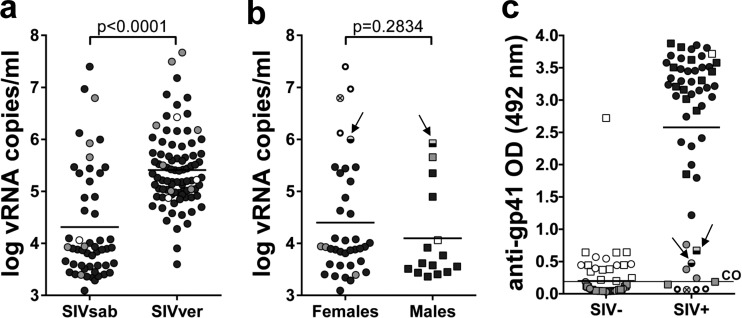
FIG 4.
Staging of SIVagmSab infection in wild sabaeus monkeys from the Gambia. (a) Significantly lower plasma viral load levels in sabaeus monkeys from the Gambia than in vervets from South Africa; (b) comparative assessment of the viral load levels between female AGMs and male AGMs from the Gambia; (c) serological testing of anti-gp41 antibodies in wild sabaeus monkeys. Infant AGMs are illustrated as open circles and squares; juvenile AGMs are illustrated as gray circles and squares; adult AGMs are shown as black circles and squares. In panels b and c, samples collected from female AGMs are illustrated as circles (○); samples collected from male AGMs are shown as squares (□); the samples from acutely infected adult females (defined as having high viral loads and negative serologies) are illustrated as open circles with enhanced margins; the sample collected from an acutely infected juvenile female is illustrated as V; finally, two samples from juvenile monkeys with high viral loads but seropositive results (probably postacutely infected) are shown as half-empty symbols and are identified by arrows. Detection limit of the VL assays, 100 copies/ml. CO, cutoff for the serological assay (arbitrarily established at 0.2) (73). OD, optical density of the serum sample.

