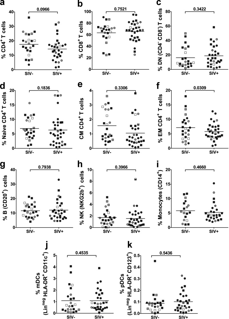
FIG 6.
Cross-sectional analysis of the major immune cell populations and subsets in wild sabaeus monkeys from the Gambia. (a) CD4+ T cells; (b) CD8+ T cells; (c) double negative (CD4− CD8−) T cells; (d) naive (CD28− CD95+) CD4+ T cells; (e) central memory (CD28+ CD95+) CD4+ T cells; (f) Effector memory (CD28+ CD95−) CD4+ T cells; (g) B cells; (h) natural killer (NKG2b+) cells; (i) monocytes (CD14+); (j) plasmacytoid dendritic cells (Linneg HLA-DR+ CD123+); (k) myeloid dendritic cells (Linneg HLA-DR+ CD11c+). Infant AGMs are illustrated as open circles and squares; juvenile AGMs are illustrated as gray circles and squares; adult AGMs are shown as black circles and squares. Samples collected from female AGMs are illustrated as circles (○); samples collected from male AGMs are shown as squares (□); the samples from acutely infected adult females (defined as having high viral loads and negative serology) are illustrated as open circles with enhanced margins; the sample collected from a juvenile male AGM with high viral loads but seropositive (probably postacutely infected) is shown as a half-empty square. P values were calculated by the Mann-Whitney U test. The values on the y axes depict the proportion of the given immune cell population or subset.

