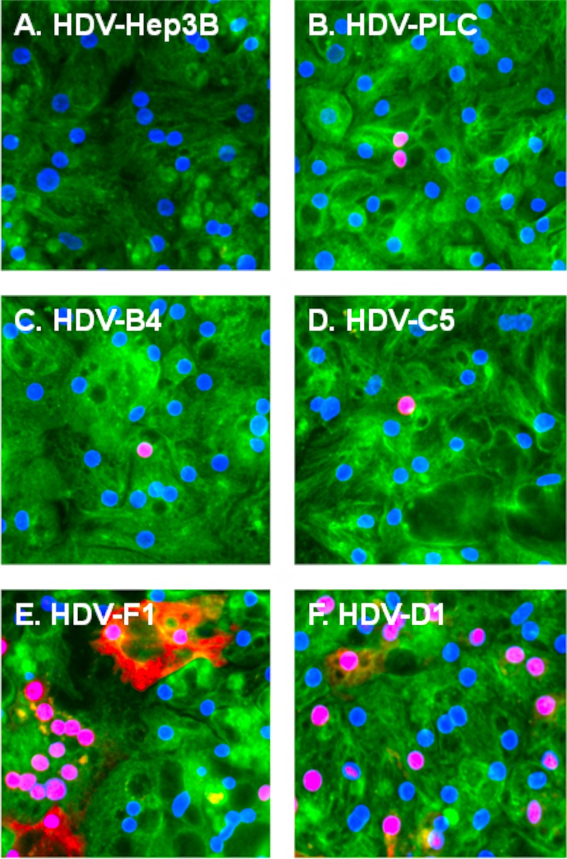
FIG 2.
Detection of PHH infected with different types of HDV virions using immunofluorescence. At day 9 postinfection, PHH were fixed, permeabilized, and analyzed for HDV-infected cells by immunofluorescence using rabbit polyclonal antibodies raised against recombinant small delta antigen (δAg) (48). Red staining is for the δAg accumulated in infected PHH. For detection of the cellular α-tubulin (green staining), mouse monoclonal antibody (Fisher Scientific) was used. Blue staining (DAPI) represents nuclear DNA. PHH were infected with HDV assembled in Hep3B cells (HDV-Hep3B) (A), HDV assembled in PLC/PRF/5 cells (HDV-PLC) (B), HDV coated with the envelope proteins of HBV genotype B, variant 4 (HDV-B4) (C), HDV coated with the envelope proteins of HBV genotype C, variant 5 (HDV-C5) (D) HDV coated with the envelope proteins of HBV genotype F, variant 1 (HDV-F1) (E), and HDV coated with the envelope proteins of HBV genotype D, variant 1 (HDV-D1) (F).

