
FIG 3.
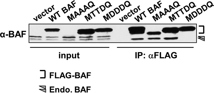
Dimerization assay of BAF by immunoprecipitation (IP). Cell lysates were subjected to immunoprecipitation using an anti-FLAG antibody followed by Western blot analyses using anti-BAF antibody. Input lysates (left) were also analyzed to illustrate the presence and migration pattern of endogenous BAF in each lysate. The bracket indicates the epitope-tagged BAF proteins, while the arrowhead indicates endogenous BAF.