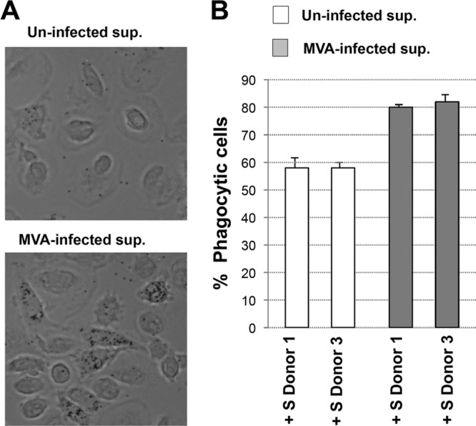
FIG 7.

MVA infection increases the phagocytosis of GFP-labeled latex beads in human macrophages. (A) Phagocytosis of GFP-latex beads by human macrophages. Cells were seeded in eight-well tissue culture plates and treated with supernatant (sup) from mock-infected (Uninfected) or MVA-infected macrophages for 16 h. After that, the cells were incubated with 1-μm-diameter latex beads conjugated to GFP in a ratio of 10 latex beads per cell. Phagocytized beads and cells were visualized 2 h after latex bead incubation by phase-contrast microscopy. Representative fields are shown at a magnification of ×40. (B) Cells having phagocytized GFP-labeled latex beads were quantified by immunofluorescence microscopy. Representative phase-contrast images are shown. The graph shows the quantification of phagocytic cells in macrophage cultures treated with supernatant (sup) from mock-infected (Uninfected) or MVA-infected macrophages. Results represent the means ± the standard deviations of three independent experiments. P values from a two-tailed t test assuming nonequal variance were determined. In all cases, P is <0.05.
