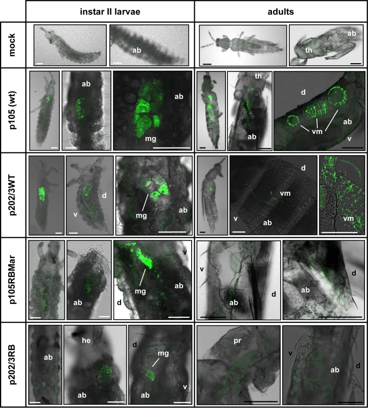
FIG 8.
TSWV infection of thrips vectors demonstrated by immunofluorescence on sections of larvae and adults. Primary antibodies were against the TSWV nucleocapsid protein. Each image is presented as the overlay of fluorescence and bright-field images. Images shown are taken from different individuals and are representative of all the observations performed (see Table 2). White bars represent 50 μm; black bars represent 100 μm. Abbreviations: ab, abdomen; d, dorsum; he, head; mg, midgut; pr, pronotum; th, thorax; v, ventre; vm, visceral muscles.

