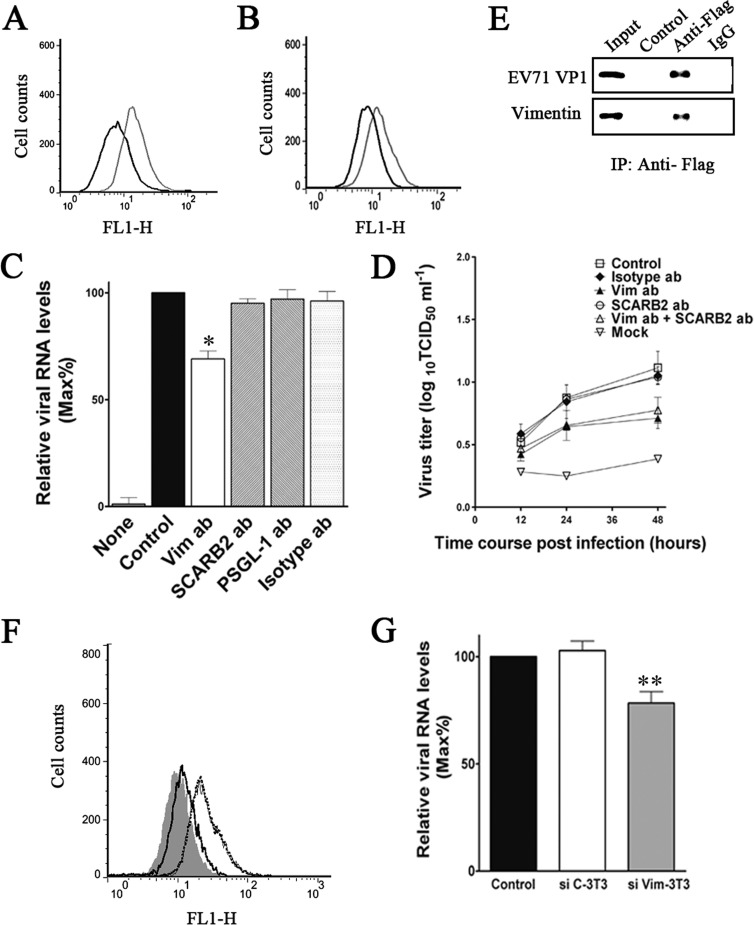
FIG 12.
Analysis of the role of vimentin in EV71 binding in mouse 3T3 cells. (A) Detection of vimentin expression on the cell surface of mouse 3T3 cells. Cells were fixed and incubated with either mouse IgG (black line) or antibody to vimentin (gray line). The cells were then incubated with the fluorescent secondary antibody and subjected to flow cytometry analysis as described in Materials and Methods. y axis (counts) = cell counts; x axis = fluorescence density. (B) Flow cytometry analysis of the binding of EV71 to 3T3 cells. Cells were incubated with EV71 at an MOI of 20 at 4°C for 1 h, fixed, and incubated with either mouse IgG (black line) or antibody to EV71 (gray line). (C) Quantitative RT-PCR analysis of the binding of EV71 to 3T3 cells that were preincubated with antibodies to vimentin, SCARB2, PSGL-1, or isotype IgG (60 μg ml−1) and then infected with EV71. Control, EV71-infected cells without preincubation; mock, uninfected cells. (D) Determination of virus titers in EV71-infected 3T3 cells and the corresponding culture supernatants at 12, 24, and 48 h postinfection. The cells were preincubated with vimentin antibody, SCARB2 antibody, or isotype IgG (control) before infection. Mock, uninfected cells. (E) Coimmunoprecipitation and Western blot analysis of the interaction between mouse vimentin and EV71 VP1 as described in Materials and Methods. 3T3 cells were transfected with plasmids expressing VP1. Coimmunoprecipitation was performed with Flag antibody. The precipitated proteins were analyzed by Western blotting using antibodies to vimentin 1 and Flag. Control, proteins from cell lysate incubated with agarose beads; VP1, proteins from cell lysate incubated with Flag-conjugated agarose beads; IgG, proteins from cell lysate incubated with IgG-conjugated agarose beads. (F) Detection of vimentin expression on the cell surface of mouse 3T3 (black dotted line), siC-3T3 (gray line), and siVim-3T3 (black line) cells. Cells were fixed and incubated with either mouse IgG (shaded area) or antibody to vimentin. The cells were then incubated with the fluorescent secondary antibody and subjected to flow cytometry analysis as described in Materials and Methods. (G) Analysis of the binding of EV71 to 3T3, siC-3T3, and siVim-3T3 cells using quantitative RT-PCR.

