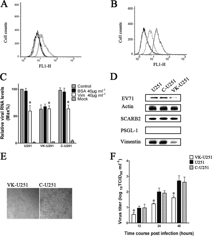
FIG 7.
Effect of the downregulation of vimentin expression in U251 cells on the efficiency of EV71 binding and replication in host cells. U251, C-U251, and VK-U251 are cells with no vector, cells with empty vector, and cells with the vimentin knockdown plasmid, respectively. (A) Flow cytometry analysis of cell surface vimentin expression in U251 (gray hairline), C-U251 (black hairline), and VK-U251 (gray heavy line) cells. Nonpermeabilized cells were fixed and stained with antibody specific to vimentin and subjected to flow cytometry analysis. Thick black line, VK-U251 cells stained with mouse IgG. y axis (counts) = cell counts; x axis (FL1-H) = fluorescence density. (B) Flow cytometry analysis of the binding of EV71 to VK-U251 and C-U251 cells. VK-U251 and C-U251 cells were incubated with EV71 for 1 h at 4°C and then fixed, stained with antibody to EV71, and subjected to flow cytometry. Black heavy line, VK-U251 cells with no EV71 infection; gray heavy line, C-U251 cells with no EV71 infection; gray dotted line, infected VK-U251 cells stained with EV71 antibody; gray hairline, infected U251 cells stained with EV71 antibody; black hairline, infected C-U251 cells stained with EV71 antibody. (C) Analysis of EV71 binding in U251, VK-U251, and C-U251 cells that had been infected with virus inoculum preincubated with vimentin before transfection by quantitative RT-PCR. Control, cells incubated with untreated EV71; BSA, cells infected with virus inoculum preincubated with BSA; Vim, cells infected with virus inoculum preincubated with vimentin; mock, cells mock treated with no virus infection. (D) Western blot analysis of EV71 binding and expression of SCARB2, PSGL-1, and vimentin in U251, C-U251, and VK-U251 cells. The respective cells were infected with EV71 at 4°C for 1 h and then lysed and blotted with antibodies to either EV71, SCARB2, PSGL-1, vimentin, or β-actin (internal control). (E) Analysis of CPE of EV71 infection in C-U251 and VK-U251 cells after infection at 37°C for 48 h using a visible light phase microscope. (F) Determination of virus titers in infected U251, C-U251, and VK-U251 cells and the corresponding culture supernatants at 12, 24, and 48 h after infection with EV71.

