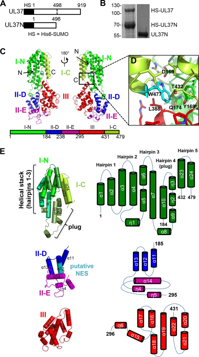
FIG 1.

UL37N structure. (A) Linear diagram of UL37 constructs expressed in this work. HS, the His6-SUMO tag. (B) Coomassie-stained SDS-polyacrylamide gel showing purified His6-SUMO-tagged UL37 contaminated with the His6-SUMO-tagged UL37N proteolytic cleavage product and purified monodisperse UL37N. (C) Crystal structure of a UL37N monomer shown in two orientations related by a 180-degree rotation around the vertical axis. (D) A close-up view of residue W477 and its surroundings. Domains are colored as in panel C. (E) UL37N domains are shown individually. The color scheme is the same as that in panels C and D, except that the layers of domain 1 are highlighted by different shades of green and the putative nuclear export signal (NES) region in domain II is shown in teal. Orientations were chosen to show all secondary structure elements. Topology diagrams displaying individual domain organization of helices are also shown.
