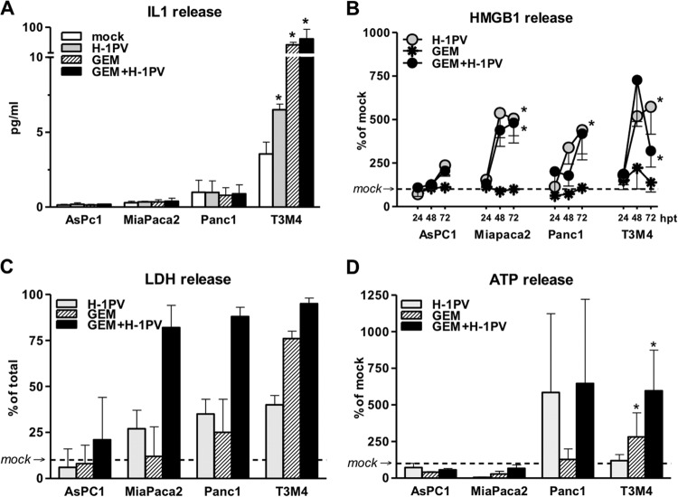
FIG 3.
Complementary induction of ICD by gemcitabine (GEM) and H-1PV in PDAC cells. PDAC cultures were treated with H-1PV at an MOI of 10 PFU/cell, with or without previous exposure to GEM for 12 h, at doses ranging from IC50 to 100× IC50 or 40 ng/ml. The supernatants were harvested between 24 and 72 hpt. (A) Selective secretion of IL-1β in T3M4 cells as determined by commercial ELISA at 48 hpt. (B) Kinetics of HMGB1 released into supernatants. The measurements are presented as percentages of levels detected in mock-infected cultures at each time point (see Fig. 1A for actual levels). (C) Levels of oncolysis in H-1PV-treated cells exposed to 40 ng/ml GEM as detected by LDH release assay at 72 hpt. (D) Selective alteration of extracellular ATP level by GEM (in relation to that in mock-infected cells) at 48 hpt. *, significantly different from mock-treated cultures (P < 0.05).

