FIG 4.
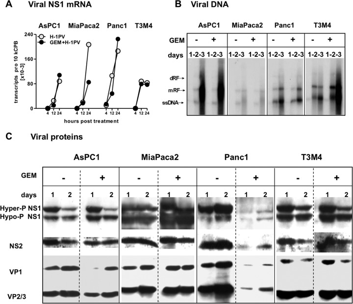
H-1PV replication in GEM-treated PDAC cells. H-1PV was added to PDAC cultures at an MOI of 10 PFU/cell, alone or after 12 hours of preexposure to GEM at the respective IC50s (see Materials and Methods). Cells were harvested at the indicated time points to measure expression of H-1PV determinants (mRNA, DNA, and proteins). (A) At 4 to 24 hpt, the number of NS1 mRNA copies was determined by qRT-PCR, normalized to cyclophilin B levels (10 kCPB), and controlled for vDNA contamination as described in Materials and Methods. (B) Viral DNA replication was assessed by Southern blotting of DNA extracts at 1 to 3 days posttreatment. ssDNA, single-stranded viral DNA genome; mRF, monomer replicative form; dRF, dimer replicative form. (C) Expression of viral proteins NS1 and -2 and VP1 to -3 was analyzed by Western blot analysis of infected cells, using antibodies targeting the respective viral proteins at 1 to 3 days posttreatment.

