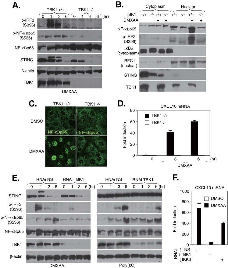
FIG 5.
TBK1 regulates DMXAA-mediated NF-κB activation in MEFs. (A) TBK1+/+ and TBK1−/− MEFs were stimulated with 200 μM DMXAA for the indicated times. The expression levels of TBK1, STING, IRF3 phosphorylated at Ser396 (p-IRF3), NF-κBp65 phosphorylated at Ser536 (p-NF-κBp65), NF-κBp65, and β-actin were determined by immunoblotting. (B) Detection of nuclear translocation of NF-κBp65 by fractionation assay. TBK1+/+ and TBK1−/− MEFs were stimulated with 200 μM DMXAA for 3 h, and then cell lysates were separated into cytosolic and nuclear fractions. Each fraction was concentrated and subjected to immunoblotting with the indicated antibodies. (C) TBK1+/+ and TBK1−/− MEFs were stimulated with 200 μM DMXAA for 3 h, and then cells were stained with antibodies against NF-κBp65. (D) TBK1+/+ and TBK1−/− MEFs were stimulated with 200 μM DMXAA for 6 h, the total RNAs were extracted from these cells, and the expression levels of mRNA for CXCL10 were determined by qRT-PCR. Data were normalized to the levels of GAPDH mRNA. (E) MEFs subjected to RNAi by use of nonspecific (NS) and TBK1 siRNAs were stimulated with 200 μM DMXAA or 5 μg/ml of poly(I·C) for the indicated times. The cell lysates were subjected to immunoblotting with the indicated antibodies. (F) MEFs subjected to RNAi by use of NS, TBK1, and IKKβ siRNAs were stimulated with 200 μM DMXAA for 6 h, the total RNAs were extracted from these cells, and the expression levels of mRNA for CXCL10 were determined by qRT-PCR. Data were normalized to the levels of GAPDH mRNA.

