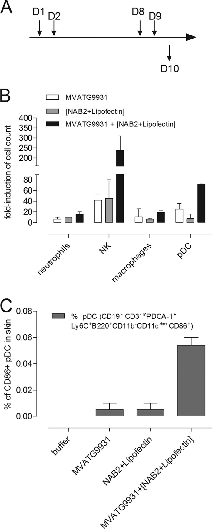
FIG 8.

Skin infiltration assay. (A) Mice were subcutaneously injected on day 1 (D1) and day 8 (D8) with 5 × 10⁵ PFU of MVATG9931. NAB2-Lipofectin (0.3 μg and 1 μg, respectively) was injected on day 2 (D2) and day 9 (D9). Mice were sacrificed on day 10 (D10). Skin around the injection site was cut out and mechanically dissociated. Suspensions of cells from 10 to 14 injection sites were immunofluorescently stained and analyzed by flow cytometry. (B) The percentages of pDCs macrophages, neutrophils, and NK within the total population were calculated, and the fold induction is expressed on the basis of the values obtained with the negative-control group (buffer injection). (C) The percentage of CD86+ pDCs in the total cell population was determined. The means ± SEMs of two independent experiments are shown.
