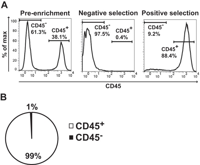
FIG 2.
LVS infects myeloid cells following intranasal inoculation. B6 mice were intranasally inoculated with 1 × 10⁴ CFU of LVS. Four hours postinfection, mice were sacrificed and lungs were removed and digested into a single-cell suspension. Cells were stained with CD45-APC, and then CD45+ cells were enriched using magnetic beads. (A) Representative flow cytometry analysis of CD45 enrichment. (B) CD45+ and CD45− populations were directly plated on chocolate agar, and the colonies were counted 72 h later. We counted 123 total CFU among 4 mice. Data are weighted by the total number of CFU and presented as the percentage of CFU within a population from 4 infected mice in 2 independent experiments.

