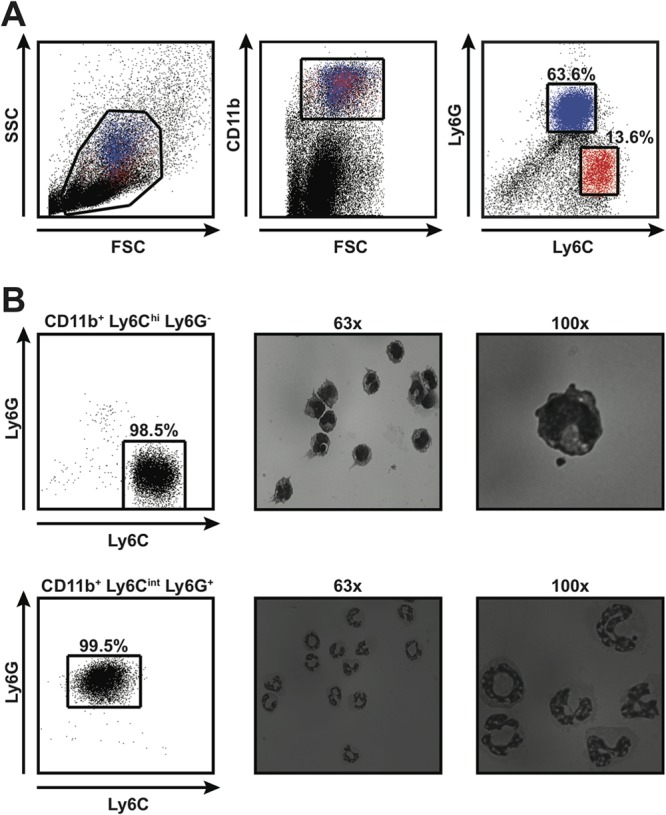
FIG 2.

CD11b+ Gr-1+ cells that accumulate in tissues of mice infected with S. Typhimurium exhibit phenotypic and morphological heterogeneity. (A) Flow cytometric analysis of splenocytes harvested from 129X1/SvJ mice infected for 28 days with S. Typhimurium. Live splenocytes (left) that expressed CD11b (middle) could be divided into CD11b+ Ly6Chi Ly6G− and CD11b+ Ly6Cint Ly6G+ cells (right). Red and blue colors indicate CD11b+ Ly6Chi Ly6G− and CD11b+ Ly6Cint Ly6G+ cells, respectively. Numbers refer to CD11b+ Ly6Chi Ly6G− and CD11b+ Ly6Cint Ly6G+ cells as percentages of live, CD11b+ cells. FSC, forward scatter; SSC, side scatter. (B) Morphological analysis of CD11b+ Ly6Chi Ly6G− and CD11b+ Ly6Cint Ly6G+ cells purified by fluorescence-activated cell sorting. The purity of the cells was consistently greater than 98% (left). Purified cells were stained with Reastain Quick-Diff and visualized by light microscopy at ×63 (middle) and ×100 (right) magnifications. Numbers refer to CD11b+ Ly6Chi Ly6G− and CD11b+ Ly6Cint Ly6G+ cells as percentages of live cells. Data are representative of six independent experiments.
