FIG 4.
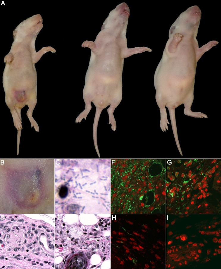
Gross pathology and histopathological examination of infant rats injected with K. kingae. The animals were i.p. injected with 2.0 × 10⁷ cells of PYKK081 or KKNB100 or with PBS and sacrificed 48 h after injection for tissue analysis. Representative pictures are shown. (A) Ventral view of the animals injected with PYKK081 (left), exhibiting an abdominal necrotic lesion and significant weight loss, KKNB100 (middle), and PBS (right). (B) Magnified view of the skin lesion formed as a result of i.p. injection of PYKK081, which demonstrated considerable necrosis, and associated acute inflammation involving the panniculus carnosus muscularis (PCM), the subdermal tissues, and presumably adherent portions of peritoneum. (C) Skin lesion section from the animal injected with PYKK081, which demonstrates muscle necrosis, some associated acute inflammation, and bacteria. Magnification, ×1,000. (D) Slide from the animal injected with KKNB100, which shows no bacteria or any histopathological changes. Magnification, ×400. (E) Skin lesion section seen in panel C. Magnification, ×400. (F to I) Immunohistochemical analysis of the skin lesion slides was performed using anti-K. kingae OMV antibody and was visualized upon interaction with Alexa 488-conjugated anti-rabbit IgG. Magnification, ×400. Nonspecific fluorescence was not observed in the absence of primary antibody. Cell nuclei were labeled with 7-AAD. An intense green signal, indicating the presence of K. kingae, was clearly observed between the abdominal thin muscle layer underlying the skin and the fatty layer in the samples from animals injected with PYKK081. (F) Skin lesion section from the animal injected with PYKK081. A mixture of intracellular organisms and extensive extracellular K. kingae was detected. (G) Lesion section from the animal injected with PYKK081. The bacteria are predominantly found inside phagocytic cells. (H) In the skin lesion section from the animal injected with KKNB100, the majority of staining was observed in phagocytic cells. The samples show much less staining. (I) Site of injection from the animal injected with PBS. No green signal was detected in these samples.

