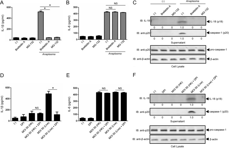
FIG 3.
Caspase-1-mediated cytokine secretion during A. phagocytophilum stimulation of macrophages is dependent on protein transport from the endoplasmic reticulum to the Golgi apparatus, the proteasome, and ROS generation. (A and B) BMDMs (1 × 10⁶) were pretreated for 1 h with brefeldin A (20 μg/ml) or MG-132 (1 μM) prior to A. phagocytophilum stimulation (MOI = 50) for 18 h. IL-1β (A) and IL-6 (B) were measured by ELISA. (C) IL-18 (p18) and mature caspase-1 (p20) in supernatants were measured by Western blotting (IB). β-Actin and pro-caspase-1 were used as loading controls. (D and E) BMDMs (1 × 10⁶) were stimulated with heat-killed (HK) or live A. phagocytophilum (MOI = 50) in the presence or absence of DPI (10 μM) for 18 h. BMDMs were pretreated with DPI for 1 h prior to stimulation. IL-1β (D) and IL-6 (E) were measured by ELISA. (F) IL-18 (p18) and mature caspase-1 (p20) in supernatants were measured by Western blotting (IB). β-Actin and pro-caspase-1 were used as loading controls. Cytokine measurements were taken in triplicate and presented as means and SEM. For panels A and B, one-way ANOVA with the Bonferroni test was used to compare nontreated and chemically treated cells. For panels D and E, Student's t test was used to compare nontreated and DPI-treated cells. (−), nonstimulated cells; *, P < 0.05; NS, not significant. Numbers below the Western blot panels show quantitation by densitometry and indicate fold differences compared to non-chemically treated samples.

