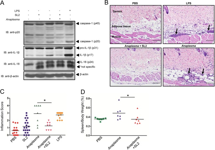
FIG 7.
Sialostatin L2 inhibits A. phagocytophilum-induced inflammation at the skin site. Intradermal injection of C57BL/6 mice was performed with PBS (−), sialostatin L2 (20 μg), A. phagocytophilum (1 × 10⁴ cells), sialostatin L2 (20 μg) plus A. phagocytophilum (1 × 10⁴ cells), and LPS (40 μg). (A) Proteins from the injection sites were extracted from skin homogenates, and Western blotting (IB) was performed to detect caspase-1 (p45 and p20), IL-1β (p31 and p17), and pro-IL-18 (p24). β-Actin was used as a loading control. (B) Skin inflammation was characterized by infiltration of neutrophils, eosinophils, and a few macrophages in the dermis, subcutaneous adipose tissue, and underlying skeletal muscle of mice (arrows). Hematoxylin and eosin staining was performed. Magnification, ×200. Bars = 100 μm. (C) Inflammation score, determined as described in Materials and Methods. (D) Spleen weights for C57BL/6 mice infected with A. phagocytophilum (n = 7) or treated with sialostatin L2 (20 μg) plus A. phagocytophilum (n = 7), normalized to the animal's body weight and contrasted to those for noninfected mice (n = 7) (PBS) at day 14 post-intraperitoneal infection. *, P < 0.05. For panel C, the Kruskal-Wallis test (with post hoc Dunn's test) was performed. For panel D, ANOVA (with post hoc Bonferroni test) was performed. NS, not significant.

