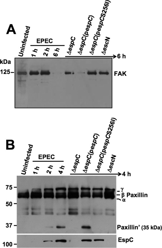
FIG 5.

EspC causes FAK and paxillin degradation. (A) FAK degradation. HEp-2 cells were infected with the strains as indicated at the top of each lane and for the indicated times. Infected cells were lysed, and proteins were analyzed by immunoblotting using anti-FAK antibody. The reaction was visualized using HRP-labeled rabbit anti-mouse antibody and developed using Western blotting chemiluminescence reagent. (B) Paxillin degradation. HEp-2 cells were infected with the strains as indicated at the top of each lane and for the indicated times. Infected cells were lysed, and the proteins were analyzed by immunoblotting using anti-paxillin as described for panel A. Note EspC detection in the cytoplasmic fraction using anti-EspC antibodies (in the same blot), indicating EspC translocation into the cells (lower panel).
