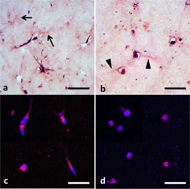
Fig. 3.

Histology of BMSCs embedded in collagen gel cultured for 7 days. HE staining of BMSCs cultured in collagen gel without (a) or with BIO (b). Arrows indicate the assembly of collagen fibers observed in the control gel, and arrowheads indicate very thin cytoplasm of round shaped BMSCs formed in BIO-supplemented gel. Immunohistochemical staining of β-catenin (red) in BMSCs cultured for 7 days in collagen gel without (c) or with BIO (d). Nuclear stainings were performed with DAPI (blue; bars=20 μm).
