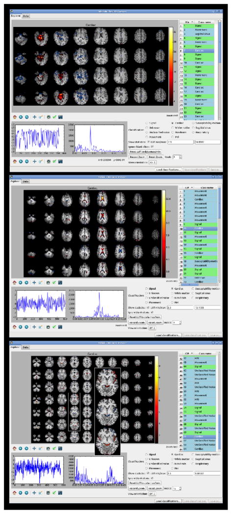
Figure 4.
Examples of cardiac-related components. This includes components due to cardiac pulsation and arterial contribution. The signal above threshold in the spatial maps is essentially located in the ventricles, or following the main arteries (posterior cerebral artery, middle cerebral branches).

