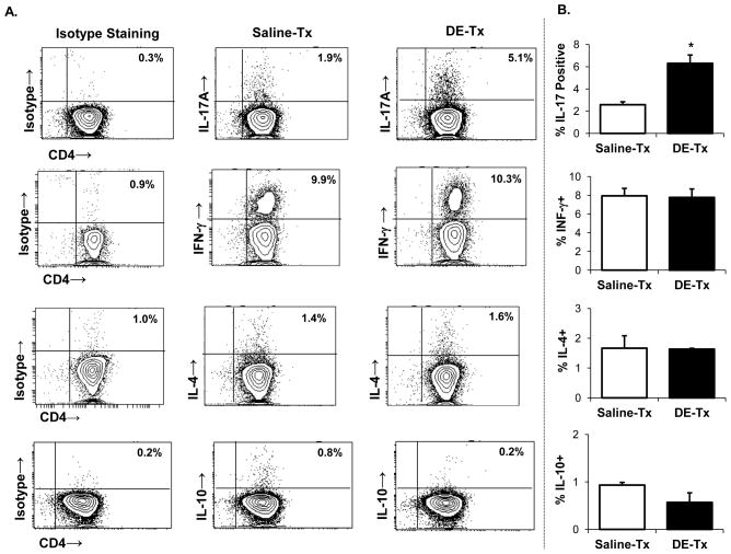
Figure 2. IL-17-producing lung CD4+ T cells are increased following repetitive DE exposure.
C57BL/6 mice were repetitively exposed to DE and saline for 3 wk whereupon CD4+ T cells pooled from three animals and isolated by FACS were immediately stimulated ex vivo with PMA + ionomycin for 4 h, and stained for IL-17 (Th17), IFN-γ (Th1), IL-4 (Th2), and IL-10 (Treg) to demonstrate cytokine profiles. A, A representative contour plot depicting cytokine staining of one of four independent experiments is shown. Left column depicts background or isotype staining for each respective cytokine. Middle and right column depict saline- and DE-treated mice, respectively. B, Results represent mean ± SEM of the percentage positive cytokine staining of CD4+ T cells with statistical difference denoted by asterisk (*p<0.05).

