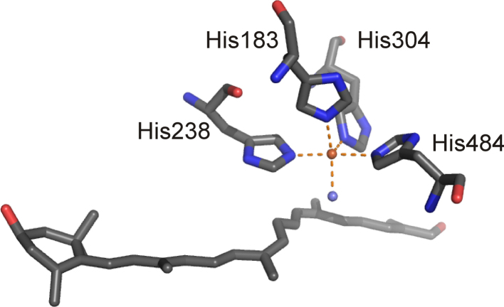
Fig. 28.
4-His metal binding site of AO (PDB: 2BIW) with bound substrate. Note that the 15-15′ double bond of the apocarotenoid substrate displays cis-configuration. Oxygen atoms and nitrogen atoms are shown in red and blue, respectively. An iron bound water molecule is shown in slate blue. The protein contains Fe(III) in the active site, which is shown in orange. (For interpretation of the references to color in this figure legend, the reader is referred to the web version of the article.)

