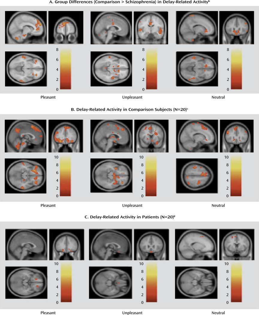
FIGURE 4. Delay-Related Brain Activity in Healthy Comparison Subjects and Patients With Schizophrenia After Viewing Pictures With Varying Emotional Valencea.
a Whole-brain statistical parametric maps of brain activity during the delay phase of an emotional experience task featuring pictures with pleasant, unpleasant, and neutral valence.
b Several of these regions fell within the predefined volume of search and survived the criterion of correction for multiple comparisons. These regions included the dorsolateral, medial, and ventrolateral prefrontal cortices for pleasant stimuli, medial frontal (supplementary motor area) and basal ganglia for unpleasant stimuli, and dorsolateral, ventromedial, and ventrolateral prefrontal cortices for neutral stimuli.
c For each type of stimulus, significant activation was generally noted in a supraset of the brain structures that showed greater activity for the healthy subjects than for the patients in the direct group contrast for the corresponding stimulus type.
d The few suprathreshold clusters identified (all shown in the figure) did not survive the correction for multiple comparisons. Uncorrected maps (p<0.05) are presented in Figure S3 in the online data supplement.

