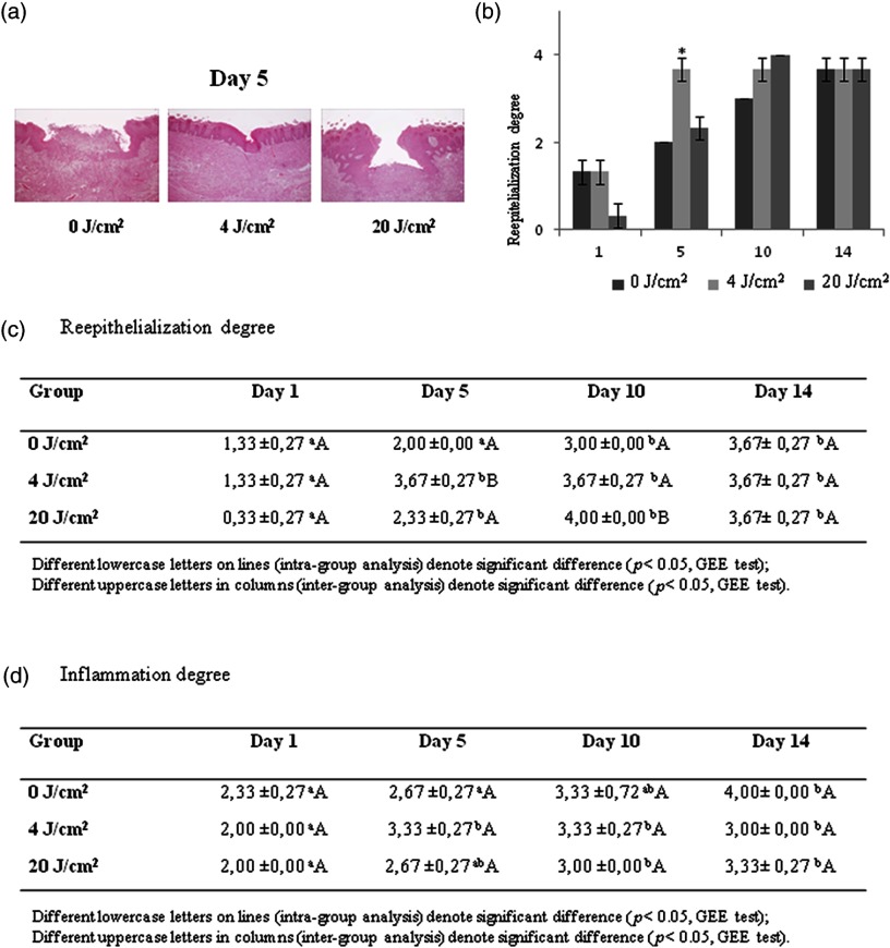
Fig. 2.
(a) Photomicrographs of experimental groups on day 5, reepithelialization covering entire wound and more chronic inflammatory infiltrate in the group [hematoxylin-eosin; magnification: (a, c, and e) and (b, d, and f)]; (b and c) Histopathological evaluation of degree of reepitelialization (mean and standard error); and (d) Histopathological evaluation of degree of inflammation (mean and standard error).

