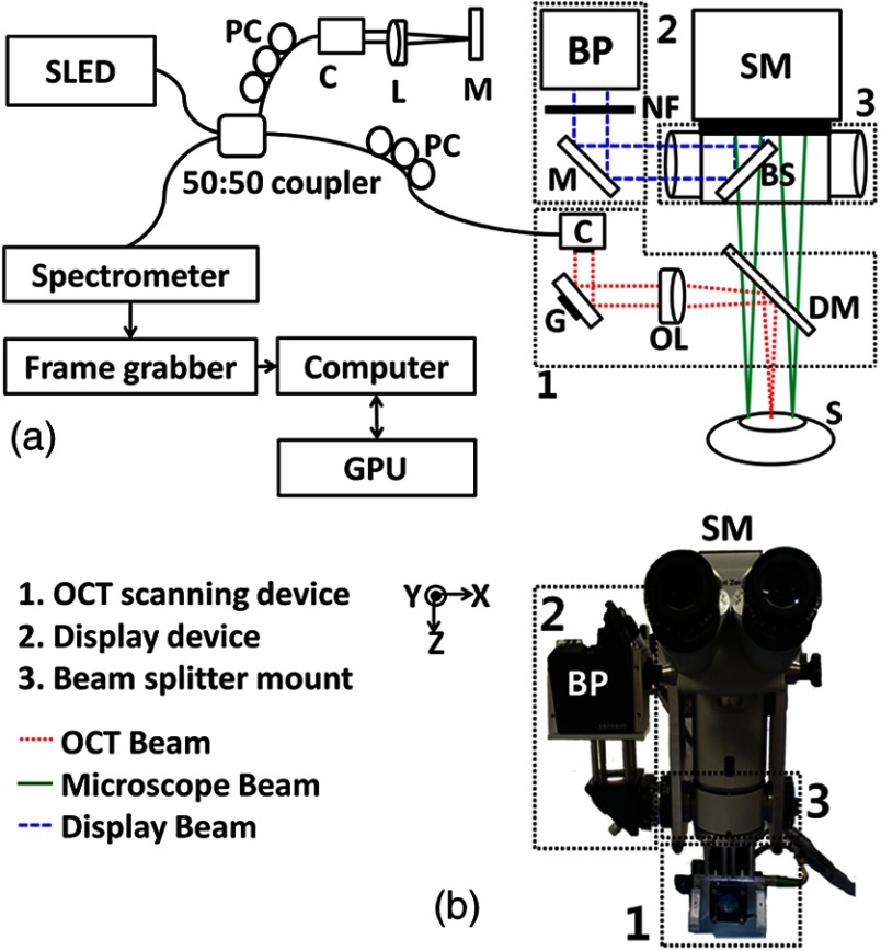
Fig. 1.
(a) Experimental setup of a real-time virtual intraoperative surgical optical coherence tomography (VISOCT) microscope. (b) Photograph of the VISOCT probe. OL, objective lens; L, lens; PC, polarization controller; DM, dichroic mirror; G, galvo scanner; BS, beam splitter; C, collimator; M, mirror; NF, neutron-density filter; BP, beam projector; SM, surgical microscope.

