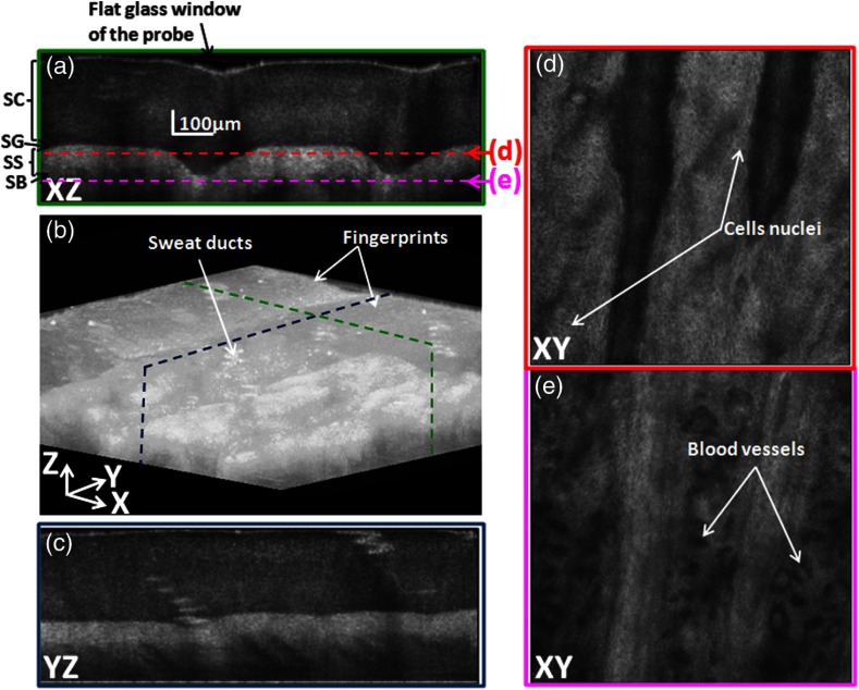
Fig. 6.
GPU-based three-dimensional (3-D) image of the pointer fingertip acquired with the architecture A (Two GTX 680); (a) and (c) represent the cross sectional images of and planes and show the different layers of the skin (SC: stratum corneum, SG: stratum granulosum, SS: stratum spinosum, and SB: stratum basale); (d) and (e) show en face images of plane at two different depths; plane (d) at around the stratum granulosum layer shows the cells nuclei whereas blood vessels can be observed on the plane (e) just below the stratum basal; (b) snap shot of the 3-D volume of 1 mm by 1 mm by 0.6 mm shows the sweat ducts.

