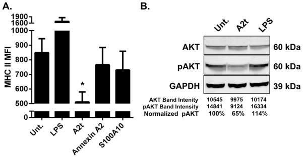
Figure 5. Exogenous A2t suppresses LC immune function.

A. LC exposed to A2t show reduced surface expression of MHC II compared to untreated LC and LC exposed to annexin A2 or S100A10. LC were left untreated (Unt.) or incubated with LPS, A2t, Annexin A2, or S100A10 and subsequently analyzed via flow cytometry for the change in expression of MHC II. The mean of three experiments ± SD is presented (*P < 0.05 as determined by a two-tailed, unpaired t-test, as compared to untreated LC). B. A2t induces an immune suppressive signal transduction cascade in LC. LC were left untreated (Unt.) or incubated with LPS or A2t for 15 min. Cellular lysates were isolated and subjected to Immuno-blot analysis demonstrating a reduction in pAKT in the A2t treated cells. One representative example of three is shown.
