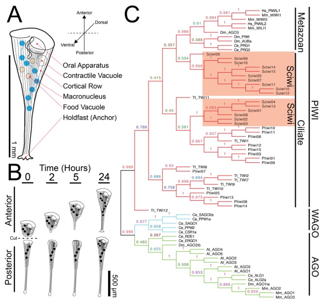
Figure 1. The anatomy of Stentor coeruleus.

(A) Cartoon of Stentor coeruleus highlighting key cellular structures, all of which have reproducible positions within the cell. (B) Cartoon representing Stentor regeneration after surgical bisection as initially reported by Thomas Hunt Morgan in 1901. (C) A neighbor-joining phylogenetic analysis of protein sequences of Stentor argonaute homologs along with sequences from Paramecium (Pt), Tetrahymena (Tt), human (Hs), mouse (Mm), C. elegans (Ce), Drosophila (Dm), and Arabidopsis (At). The three major classes of Argonaute proteins—PIWI, WAGO, and AGO—are indicated (Sciwi proteins listed in Table S1, gene IDs in Table S2).
