. 2014 May 13;12(5):e1001862. doi: 10.1371/journal.pbio.1001862

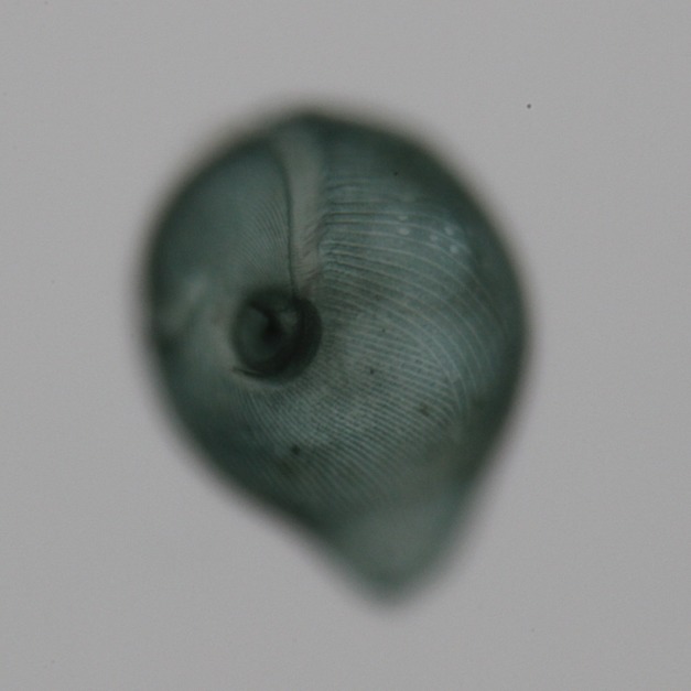
Stentor coeruleus . Brightfield image of Stentor coeruleus clearly shows the cell's stripes. The feeding organelle, or oral apparatus, is present at the top of the image while the cell anchor is oriented toward the bottom of the image. Image credit: Mark M. Slabodnick - University of California, San Francisco.

Stentor coeruleus
. Brightfield image of Stentor coeruleus clearly shows the cell's stripes. The feeding organelle, or oral apparatus, is present at the top of the image while the cell anchor is oriented toward the bottom of the image. Image credit: Mark M. Slabodnick - University of California, San Francisco.