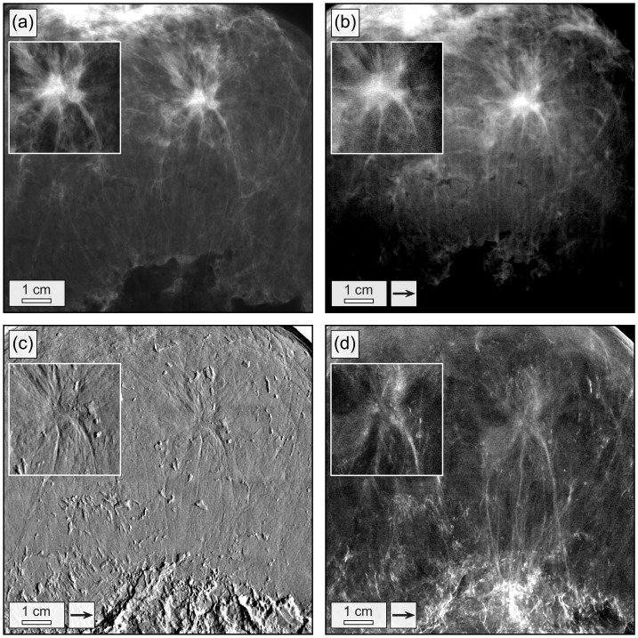
Figure 2. Conventional and grating-based mammograms of a breast specimen with invasive ductal carcinoma.
Conventional mammogram (a), grating-based absorption (b), differential phase (c) and dark-field image (d) of the breast specimen. The field of view has a size of 12.8×12.8 cm2. Images (b)–(d) were obtained by stitching together 4×4 low-statistic projections. Arrows indicate the direction of scanning. The high-statistic images of the invasive ductal carcinoma are shown as inlays.

