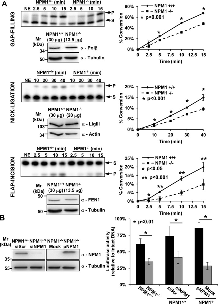
FIGURE 3:
NPM1 contributes to the tuning of BER capacity. (A) In vitro BER assays using NPM1+/+ and NPM1−/− whole-cell extracts at equal amounts of DNA Polβ, DNA ligase III (LigIII), or FEN1 reveal impaired gap-filling, nick-ligation, and flap-incision activities, respectively, in the absence of NPM1. For each enzymatic activity we report a representative denaturing gel analysis, along with a Western blot indicating the amount of cell extract used to obtain comparable quantities of each BER factor (left). Graphs (right) describe the percentage of substrate (S) converted to product (P) as a function of time. Values reported are mean ± SD of at least three independent experimental replicates. (B) In vivo BER capacity assessment using a reporter-based system as indicated in Materials and Methods (left). The assays highlight a positive role for NPM1 in the modulation of BER capacity. Histograms show mean ± SD of at least three independent replicates. Luciferase activity for the depurinated plasmid relative to that of undamaged DNA is reported. A statistically significant impairment in BER capacity is observed when comparing NPM1+/+ and NPM1−/− cells. A similar defect in BER-mediated repair is also visible when comparing siScr- and siNPM1-treated NPM1+/+ cells. Reconstitution of NPM1−/− cells with human recombinant NPM1 (pNPM1) instead restored the BER defect. A representative Western blotting analysis (right) shows NPM1 levels in the siScr- and siNPM1-treated NPM1+/+ cells and in the mock- and NPM1-reconstituted (pNPM1) NPM1−/− cells used in the in vivo BER assays. Tubulin was used as loading control.

