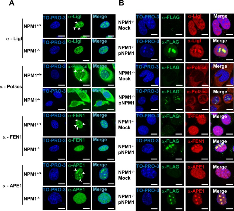
FIGURE 4:
NPM1 promotes the accumulation of BER proteins within nucleoli. (A) Representative immunofluorescence images showing the differential subcellular localization of diverse BER factors in NPM1+/+ and NPM1−/− cells. Each protein analyzed (left) shows nucleolar accumulation (white arrowheads) only in the presence of NPM1. TO-PRO-3 counterstaining was used to highlight nuclei; bars, 16 μm. (B) Representative immunofluorescence analysis on mock- and NPM1-reconstituted (pNPM1) NPM1−/− cells. NPM1 staining (α-FLAG, green) was used to localize cells positively transfected with FLAG-tagged human NPM1; NPM1 reexpression restores the accumulation of BER proteins (red) within nucleoli. Nuclei are counterstained with TO-PRO-3; bars, 16 μm.

