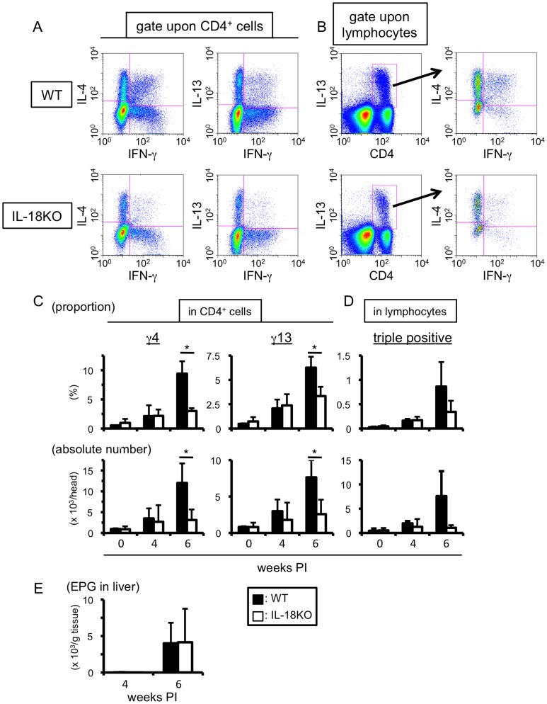
Figure 3. IL-18 plays a role in the expansion of MCPHT cells during S. mansoni infection.
(A–D) Hepatic lymphocytes were isolated from wild type (WT) or IL-18-deficient (IL-18KO) mice at indicated time points of S. mansoni infection, and the proportions and absolute numbers of γ4, γ13 (A and C), or triple positive (B and D) cells were analyzed by ICS upon TCR ligation. (A and B) One example using hepatic lymphocytes prepared at 6 weeks PI was exhibited. (C and D) Upper graphs; the percentages express the proportions in CD4-positive (γ4 or γ13 cells, C) or in lymphocyte (triple positive cells, D) population. Lower graphs; the absolute numbers of γ4, γ13 (C), or triple positive (D) cells were demonstrated. Data are expressed as mean values+SD of three or four mice in each experimental time point. Data shown are a representative of three independent experiments. (C) *0.02<P<0.05 (Mann-Whitney U test). (E) EPG of WT and IL-18 KO mice were analyzed at 4 and 6 weeks PI. Data represent the mean values+SD of four mice in each experimental time point. This is one representative of three independent experiments. (C–E) Open bars represent WT mice, and filled bars do IL-18KO mice.

