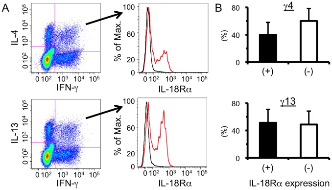
Figure 4. IL-18 receptor is expressed upon some MCPHT cells.
(A) Hepatic lymphocytes were isolated from S. mansoni-infected mice at 6 weeks PI and ICS was conducted after TCR ligation. The expression levels of IL-18 receptor α (IL-18Rα) upon γ4 (upper panels) or γ13 (lower panels) cells were analyzed. Cells were stained with anti-IL-18Rα (red line) or its respective isotype control (black line). One representative result is shown. (B) The proportions of IL-18Rα-positive (filled bars) or -negative (open bars) population in γ4 (upper graph) or γ13 (lower graph) cells are shown. Data shown are a representative of four independent experiments.

