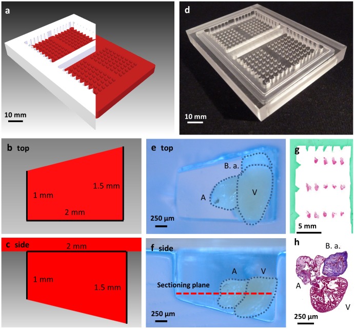
Figure 3. Heart mould for high throughput histology.
(a) 3D render of cutaway top (white) and bottom part (red) of the designed heart mould. (b) Render of single well from above with superimposed dimensions. (c) 2D cutaway of a single well of the heart mould showing dimensions. (d) Top and bottom sections of heart mould machined from acrylic. (e) Top down view of single agar well with zebrafish heart. (f) Cut-away side view of single well showing heart and sectioning plane. (g) Example slide with grid of hearts with histology-dye borders showing arrow row and column markers. (h) High-magnification of single H&E stained heart. V, ventricle. A, atrium. B.a., bulbus arteriosus.

