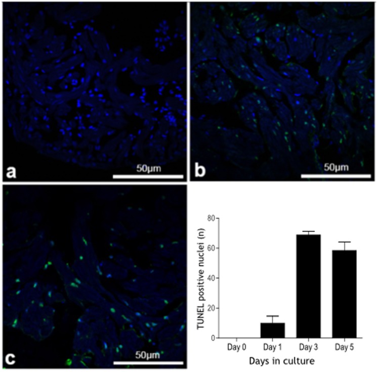
Figure 6. Apoptosis in isolated cultured zebrafish hearts (TUNEL staining).
Apoptotic nuclei (green) and nuclear DAPI staining (blue) in zebrafish hearts (assessed by TUNEL staining) maintained in culture for 0, 1, 3 and 5 days showing a marked increase in the number of apoptotic bodies at day 3 which is maintained at day 5.

