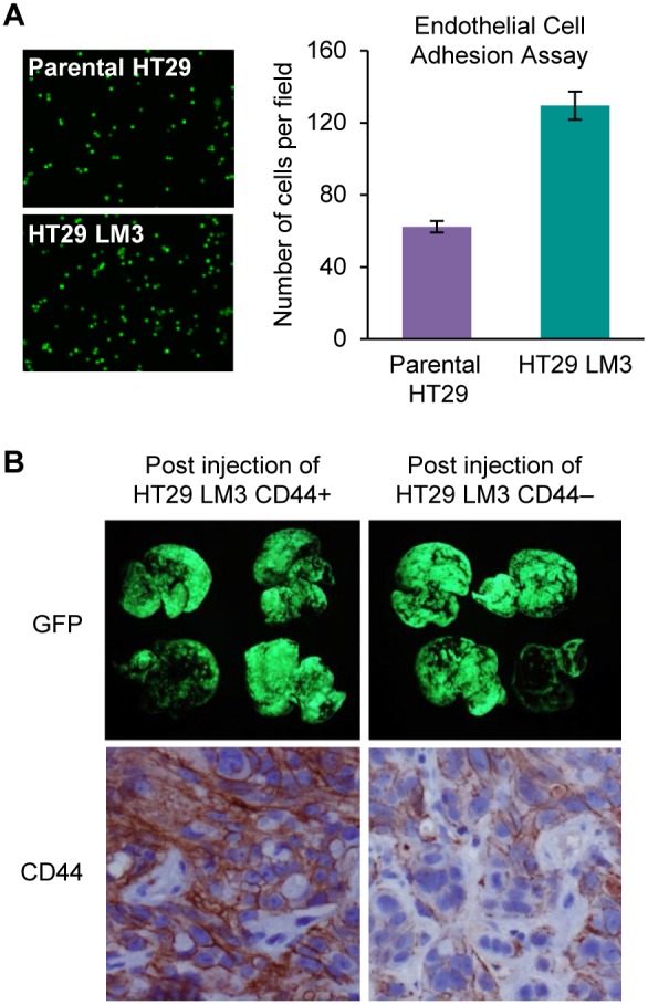
Figure 5. Role of CD44 high and CD44 low expression in CRC liver metastasis.

(A) Adhesion of parental HT29 and HT29 LM3 cells to HMVEC-L cells was assessed as described in Materials and Methods. Data shown as mean fold changes in number of parental HT29 cells attached to HMVEC-L cells versus HT29 LM3 cells (*p<0.001). Representative images show adhesion of parental HT29 and HT29 LM3 cells to HMVEC-L cells. (B) Fluorescent GFP imaging of liver metastasis 4 wks after intrasplenic injection of HT29 LM3 CD44+ and HT29 LM3 CD44− cells (5×10⁶; 100 µl of PBS). IHC analysis of CD44 expression in CD44 high and CD44 low CRC liver metastasis.
