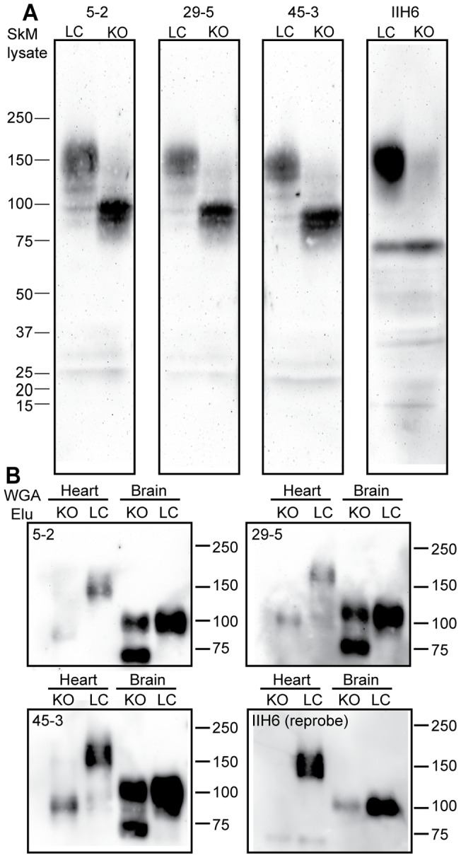
Figure 1. Detection of αDG core protein from skeletal muscle of normal and dystroglycanopathy mice.

A) Western blot analysis of αDG core protein detection by rabbit aDGct supernatants. Monoclonal antibodies 5–2, 29–5, and 45–3 were tested on replicate Western blots of solubilized murine skeletal muscle. Lane 1 contains normal murine skeletal muscle (LC) and lane 2 contains skeletal muscle from a mouse with a tamoxifen-induced fukutin-deficient dystroglycanopathy (KO). Detection with antibody IIH6 shows glycosylated αDG for comparison. Molecular weight standards are indicated in kDa. B) Wheat germ agglutinin (WGA) purifications of LC and KO mouse brain and heart lysates were conducted and the elution fraction was analyzed by Western blot. Monoclonal antibody media supernatants 5–2, 29–5, and 45–3 were used to detect αDG core protein on replicate blots; detection with IIH6 was performed by reprobing stripped blots. Molecular weight standards are indicated in kDa.
