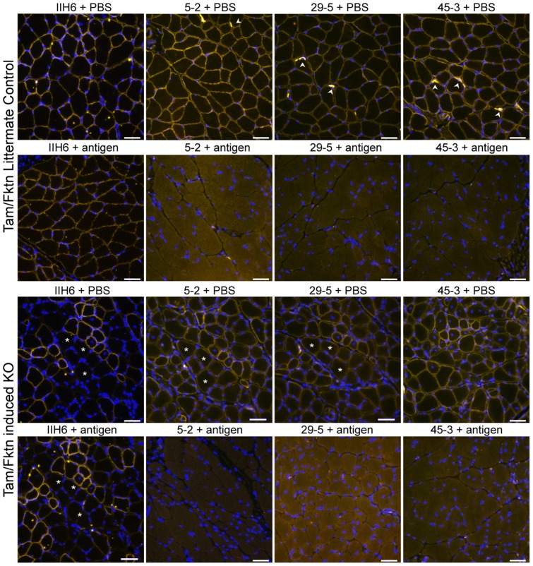
Figure 2. Immunofluorescent detection of αDG core protein on muscle sections of normal and dystroglycanopathy mice in the presence and absence of the aDGct antigen.
Calf muscle cryosections from a normal mouse (LC) and a tamoxifen-induced fukutin-deficient dystroglycanopathy mouse (KO) were stained by αDG core monoclonal antibody media supernatants 5–2, 29–5, and 45–3, as well as IIH6, for detection of αDG protein and functional glycan, respectively. Each antibody was pre-incubated with 18 µg of purified aDGct protein antigen or PBS prior to staining. Asterisks mark matching fibers for comparison. Arrowheads mark neuromuscular junctions. 40X objective; 20 µm scale bar.

