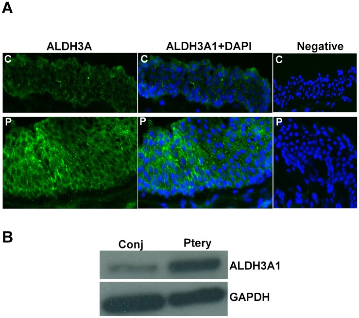
Figure 1. ALDH3A1 expression in pterygium.
A. Protein expression of ALDH3A1 in both conjunctiva and pterygium fibroblast cells was determined by immunoblotting with antibodies specific for ALDH3A1 and GAPDH. B. Immunofluorescent staining image showing presence of ALDH3A1 (green) in human conjunctiva and pterygium epithelium. Nuclei were stained with DAPI (blue) present in the mounting medium. Negative control (without primary antibody) was shown. All images were taken at 200× magnification. C: conjunctiva; P: pterygium.

