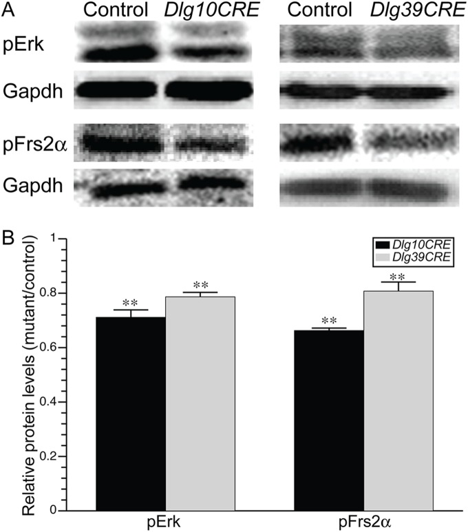
Figure 2. Levels of Fgfr signaling intermediates are reduced in both Dlg10CRE and Dlg39CRE fiber cells.
(A) RIPA lysates from P2 control, Dlg10CRE, and Dlg39CRE fiber cells were immunoblotted for the pErk and pFrs2α and the blots reprobed for Gapdh as a loading control. Representative blots are shown. (B) Quantification of protein levels. Shown are the levels of the indicated proteins in extracts from Dlg10CRE fiber cells relative to levels in the controls (control levels set at 1.0). Signal intensities were quantified by phosphorimager analysis, as described in Materials and Methods, and the data subjected to statistical analysis using the two-sided One Sample t-test. At least 3 protein pools were blotted in triplicate over 1–3 blots. The relative levels of Fgfr signaling intermediates were reduced in the fiber cells of Dlg39CRE mice as well as Dlg10CRE mice compared to controls, indicating that the effect was fiber cell autonomous. Error bars = standard deviations. ** = FDR<0.01.

