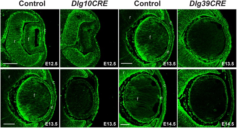
Figure 4. Loss of Dlg-1 protein following cre-mediated excision of Dlg-1 sequences.
Paraffin embedded sections of eyes from control, Dlg10CRE and Dlg39CRE day E12.5, E13.5, and E14.5 embryos were subjected to immunoflourescent staining for Dlg-1 (green). For Dlg10CRE lenses, the intensity of staining throughout the lens was greatly reduced at E12.5 and staining was undetectable at E13.5. For Dlg39CRE lenses, the intensity of staining in the fiber cell compartment was greatly reduced at E13.5 and staining in the fiber cell compartment was undetectable at E14.5. c, cornea; e, lens epithelium; f, lens fiber cells; r, retina. Bar = 50 µm.

