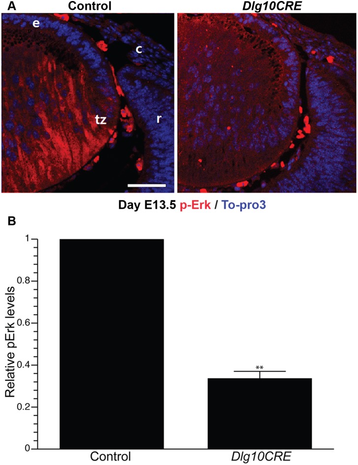
Figure 5. Levels of the Fgfr signaling intermediate, pErk, are reduced in embryonic Dlg10CRE lenses.
(A) Paraffin embedded sections of eyes from E13.5 control and Dlg10CRE embryos were subjected to immunofluorescence analysis using an anti-pErk antibody (red) and the nuclei counterstained with To-Pro3 (blue). Representative images of the transition zone are shown. c, cornea; e, lens epithelium; r, retina; tz, transition zone. Bar = 50 µm. (B) Quantification of pErk levels. Shown are the relative levels of pErk in the transition zone of DLG10CRE and control lenses (control levels set at 1.0) Quantification of signal intensities in the transition zone was carried out using ImageJ, as described in Materials and Methods, and the data subjected to statistical analysis using the two-sided One Sample t-test. At least 3 different sections from at least 3 different lenses were evaluated. The relative levels of pErk in the transition zone of the Dlg10CRE lens were reduced as compared to levels in the corresponding region of the control lenses. Error bars = standard deviations. * = FDR<0.01.

