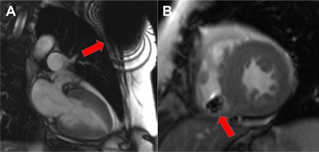
Figure 1.

Balanced steady-state free precession (b-SSFP) cine images of the heart in a patient with an implanted magnetic resonance imaging-conditional pacemaker.
Notes: Note the susceptibility artifacts from the pulse generator in left ventricular 2-chamber view (A) and from the right ventricular lead in short-axis view (B). Despite being clearly apparent, these artifacts don’t usually hinder diagnostic interpretation, except when the region of interest is in the proximity of the pacemaker generator.
