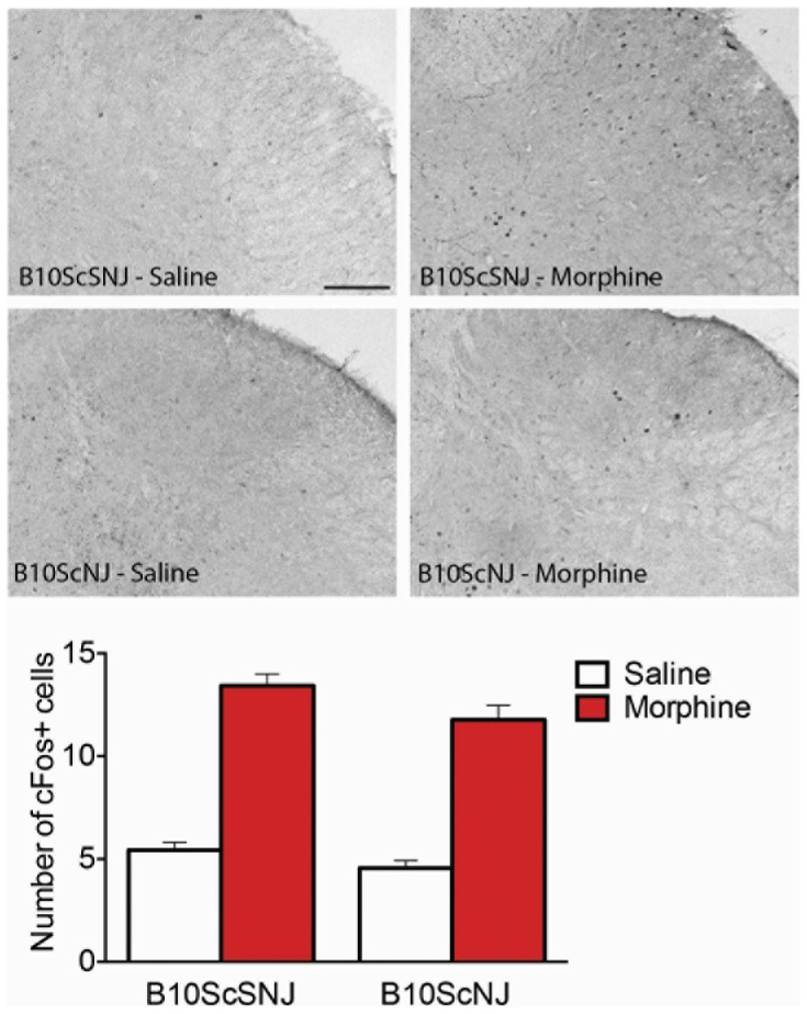
Figure 6. c-Fos immunohistochemical labeling in the dorsal spinal cord following naloxone precipitated withdrawal in morphine treated control and TLR null mice.
Mice underwent cardiac perfusions with 4% PFA 2 h following naloxone precipitated withdrawal and spinal cords were processed for immunohistochemistry of c-fos labeling. Significant differences in labeling intensity were produced by morphine in both control (B10SCSNJ) and mutant (B10ScNJ) mice. Data represent means for n = 5–6 per group. Statistical analyses were performed using a two-way ANOVA followed by Bonferroni post hoc test. p>0.05 for genotype.

